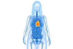
Causes Diagnosis and Treatment of Chronic Constrictive Pericarditis
Meaning of Chronic Constrictive Pericarditis: Subacute exudative
constrictive pericarditis refers to a special type of lesion in which the
visceral layer of the pericardium is narrowed with a large amount of
pericardial exudate.
It can be caused by tuberculosis, recurrent nonspecific
pericarditis, trauma, radiation, rheumatoid arthritis, uremia, and scleroderma.
Some reasons are unknown. The clinical manifestations are both pericardial
tamponade and constriction. Qimai is more common than constrictive
pericarditis. The X-ray indicates that the heart shadow is obviously enlarged,
and pericardial calcification is rarely seen.
After injection of gas into the
pericardium, the pericardial wall thickened and the heart shadow was normal or
reduced. ECG diagram QRS wave low voltage, T wave low or inverted, after
pericardial puncture and drainage, the central venous pressure and right atrial
pressure remain at the original high level.
The right atrial pressure curve has
obvious X tilt or Y tilt equal.
After the liquid is drained, Y tilts deeper.
The disease often progresses to constrictive pericarditis within one year.
Although
treatment with hormones and pericardial puncture can achieve temporary results,
it cannot prevent it from developing into constrictive pericarditis, so
treatment mainly depends on pericardial dissection.
It is a disease caused by thickening, adhesion and even
calcification of the pericardium caused by chronic inflammation of the
pericardium, which restricts the relaxation and contraction of the heart,
reduces cardiac function, and causes disorders of systemic blood circulation.
Pericarditis Reference
1. Etiology and pathology
Chronic constrictive pericarditis
is mostly caused by tuberculous pericarditis. Acute suppurative pericarditis is
about 10% of those who do not heal, and others can also be caused by
rheumatism, trauma, and mediastinal radiotherapy.
Pathological changes occur in the pericardial wall and the
visceral layer. As the lesion progresses, the pericardium adheres, thickens,
and even calcifies.
The generally thickened pericardium binds the heart. A
narrow ring can be formed at the entrance of the vena cava, causing severe
obstruction, and severe narrowing in the atrioventricular groove, causing
patients to have symptoms and signs similar to atrioventricular valve stenosis.
Due to limited cardiac activity, the myocardium undergoes disuse atrophy early,
and myocardial fibrosis can occur later.
Due to apparently limited diastole,
reduced filling volume, weakened myocardial contractility, increased
ventricular diastolic pressure, restricted venous return, and increased venous
pressure, congestion of various organs throughout the body, jugular venous
dilatation, hepatomegaly, ascites, and pleural effusion Wait for signs.
2. Clinical manifestations of Pericarditis
Tuberculous pericarditis can
appear symptoms 3 to 6 months after the acute phase. The common ones are
fatigue, shortness of breath, oliguria, abdominal distension, loss of appetite,
ascites, and hepatomegaly, which lead to systemic edema.
3. Physical examination of Pericarditis
Those with narrowing and heavier
constriction usually present with chronic disease, superficial neck veins
dilate, apical beats weaken or disappear, fast heart rate, weak heart sounds,
fine pulse and odd pulse.
The abdomen is swollen and frog-shaped, the liver is
enlarged, and the liver neck sign is positive. Blood pressure is at a low
level, and the pulse pressure difference narrows. The central venous pressure
can rise above 20cmH2O.
4. Auxiliary inspection for Pericarditis
X-ray: the heart shadow is normal or slightly enlarged, the
left and right heart borders are straightened, the superior vena cava shadow is
widened, the heart beat is weakened, and there may be signs of pericardial
calcification or pleural effusion.
Electrocardiogram: QRS complex low voltage in each lead, T
wave low or inverted. Atrial fibrillation can be seen in some patients.
Echocardiography: pericardial thickening, adhesions, effusion
and calcification, enlarged atria, ventricular contraction, and decreased
cardiac function.
Right heart catheterization: cardiac output is lower than
normal. The pressure in various parts of the heart cavity is generally
increased, the pulmonary capillary pressure is also increased, the right
ventricular diastolic pressure is significantly increased, the early diastolic
sagging, and the late increase.
5. Differential diagnosis
Chronic constrictive pericarditis
is not difficult to diagnose based on medical history, symptoms and physical
examination, but it is often necessary to distinguish between heart failure
caused by cardiomyopathy, cirrhosis, and valvular disease.
6. Surgical Treatment of Pericarditis
Once the diagnosis of chronic
constrictive pericarditis is confirmed, surgical treatment should be performed
as soon as possible.
Before the operation, make preparations according to the
patient's condition. Preparations should be like limiting sodium salt, proper application of
diuretics, maintaining water and electrolyte balance, strengthening nutrition,
supplementing protein, vitamins, small amounts of blood transfusion or plasma,
anti-tuberculosis treatment for tuberculosis patients, and appropriate elimination
of pleural effusion and ascites.
The surgical approach often uses longitudinal sternal
approach or left anterior thoracic incision. Make a small incision in the
thickened pericardium in front of the ventricle. Be careful when approaching
the myocardium.
When the pericardium wall and the visceral layer are between,
the myocardium bulges slightly when the heart contracts, and the wall and the
visceral layer are up and down and both sides Separation, the separation
sequence is the left ventricle, then the right ventricular outflow tract, the
right ventricular base, and finally the superior and inferior vena cava are
released.
Both sides of the pericardial resection range should reach the
phrenic nerve, up to the root of the aorta, and down to most of the phrenic
surface. Prevent damage to coronary arteries and myocardium during separation.
In critically ill patients, the surgical mortality rate is
relatively high. About 75% of deaths are due to acute or subacute heart
failure. Therefore, strict postoperative root volume and appropriate cardiac
support are still an important part of ensuring successful surgery.
Author's Bio
Name: Gwynneth May
Educational Qualification: MBBS, M.D. (Medicine) Gold Medalist
Profession: Doctor
Experience: 16 Years of Work Experience as a Medical Practitioner


Comments
Post a Comment